Ortho Tx: What Happens with Ortho Tx?

2. Harmony between the temporomandibular joints (TMJs) and tooth fit

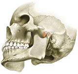
There is a direct relationship between tooth fit and the TMJs. As shown in the figure, the mandible has teeth in the middle of the bone, and the ends (condyles) are seated in the joints. Harmonizing the TMJs and tooth fit is one of the important missions of orthodontic treatment.

A TMJ problem may be associated with joint sound, difficulty opening the mouth or TMJ pain. However any of these symptoms may be caused by other factors than tooth fit, so differential diagnosis is needed.

With children, TMJ problems have direct effect on jaw growth, and such cases therefore must be diagnosed with care.

What is a good harmony between the TMJs and tooth fit?
the temporomandibular joints the temporomandibular joints
in harmony
out of harmony
out of harmony
Importance of TMJs as revealed by experiments on animals
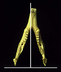
Lower jaw bone of animal

When purposely creating a positional shift of the disc in animals' right TMJ, lower jaw growth is limited on the right side. This results in a displacement of the lower jaw's midline toward the right, as seen in the figure. An analogy would be driving a car and stepping onto a brake that only works on the right side; the car will instantly spin to the right. When this happens, the right side of the lower jaw is not only shorter in length, but is also suppressed vertically in its growth. As this experiment demonstrates, TMJ problems greatly affect lower jaw growth.
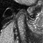
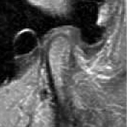
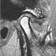
TMJs and discs

Now, let's take a look at the mechanism of TMJs through patients' MRI and CT images.

Optimum TMJ
Optimum TMJ
MRI: The convexity on the rear side of the disc is positioned at the 12 o'clock position. / CT image: The condyle, which is a part of the lower jaw, is located within the eminence in upper and slightly forward position.
TMJ disc slightly displaced forwards
MRI: This is a 12 years old patient with a disc displaced to the front. The lower jaw position within the TMJ consequently shifts backwards, and the midline moves to the right. Simultaneously, molars on the right side begin to excessively grind together, eventually wearing out the teeth. / CT image: Lower jaw's condyle is positioned slightly toward the rear, and structural change is observed at the tip of the lower jaw bone.
TMJ disc displaced significantly
MRI: The disc is completely displaced forward and downward. The jaw may get stuck, and the patient may feel some pain while trying to open the mouth. One common symptom observed in patients before this stage of progress in TMJ problems is the clicking noise at the joints. With dislocated discs, bone structure at the joints will change, and the lower jaw begins to shift significantly. The bite relationship will be lost. / CT image: The condyle is further displaced upward and toward the rear, and the bone structure has also changed. Likewise, the shape of the articular fossa and eminence will change.